A Sinus Lift Surgery is a procedure performed in the upper jaw or "Maxilla" in the region directly adjacent to the Maxillary Sinus, commonly to augment/replace the missing bone required for Dental Implant placement.
After extraction of teeth close to the floor of the maxillary sinus, surgeon sometimes encounter lack of adequate bone to successfully place a Dental Implant to replace the missing teeth. This happens as a result of a combination of natural reduction in bone volume at the extraction site and an increase in the sinus volume.
In such cases, an Oral Surgeon performs a "Sinus Lift" surgery, to add bone in a space created by meticulously handling the sinus membrane.
This surgery can be performed under Local Anesthesia or under IV Sedation/General Anesthesia.
Bone graft requires 4-6 months of healing. Sometimes Dental Implants can be simultaneously placed during the sinus lift surgery, and your Oral Surgeon can discuss with you, about this option during the consultation.
Patients are placed on Antibiotics and Pain Medications after the surgery. Surgeons will provide specific post surgical instructions related to the sinuses. Pain and discomfort is usually well controlled and may last 48-72 hours. Some patients may experience increased sinus drainage in the immediate post surgical period.
The following pictures and video, will help in better understanding the surgical procedure. The surgery is performed by Dr Abhishek Mogre. The patient has reduced bone volume in upper left first molar, and was treated with Sinus Lift Surgery to replace the missing bone in preparation for a Dental Implant.
This 3D image shows lack of bone in the missing maxillary left first molar area and enlarged "Pneumatized" Maxillary sinus.
Bony defect in the Maxillary missing first molar area, seen as a concavity.
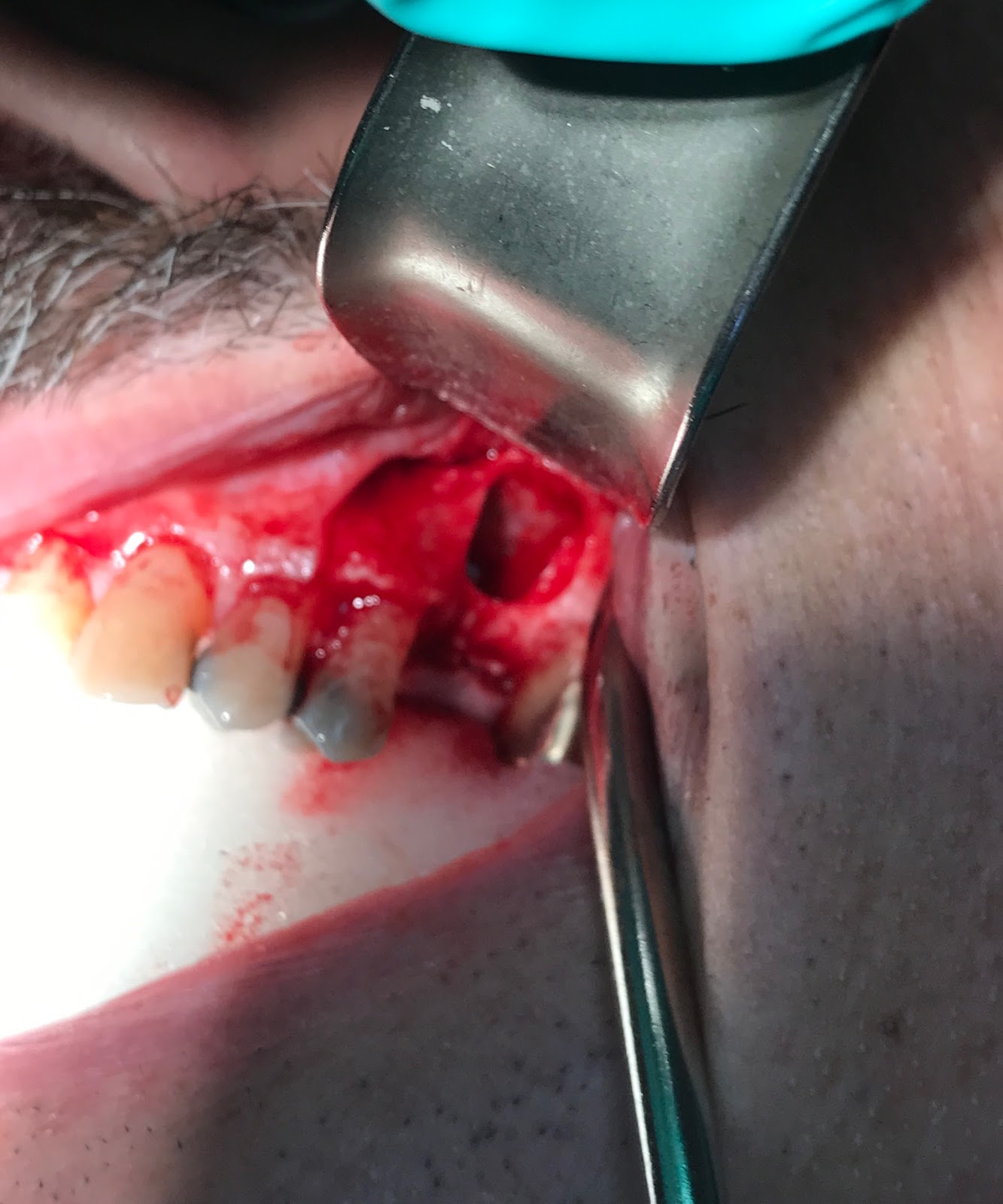
Surgical exposure of the defect
Access made to the Sinus Membrane with a window in the thin Bone in the area of the missing tooth. At this point, Sinus Membrane has been carefully manipulated.
The video shows movement of the manipulated sinus membrane during normal breathing.
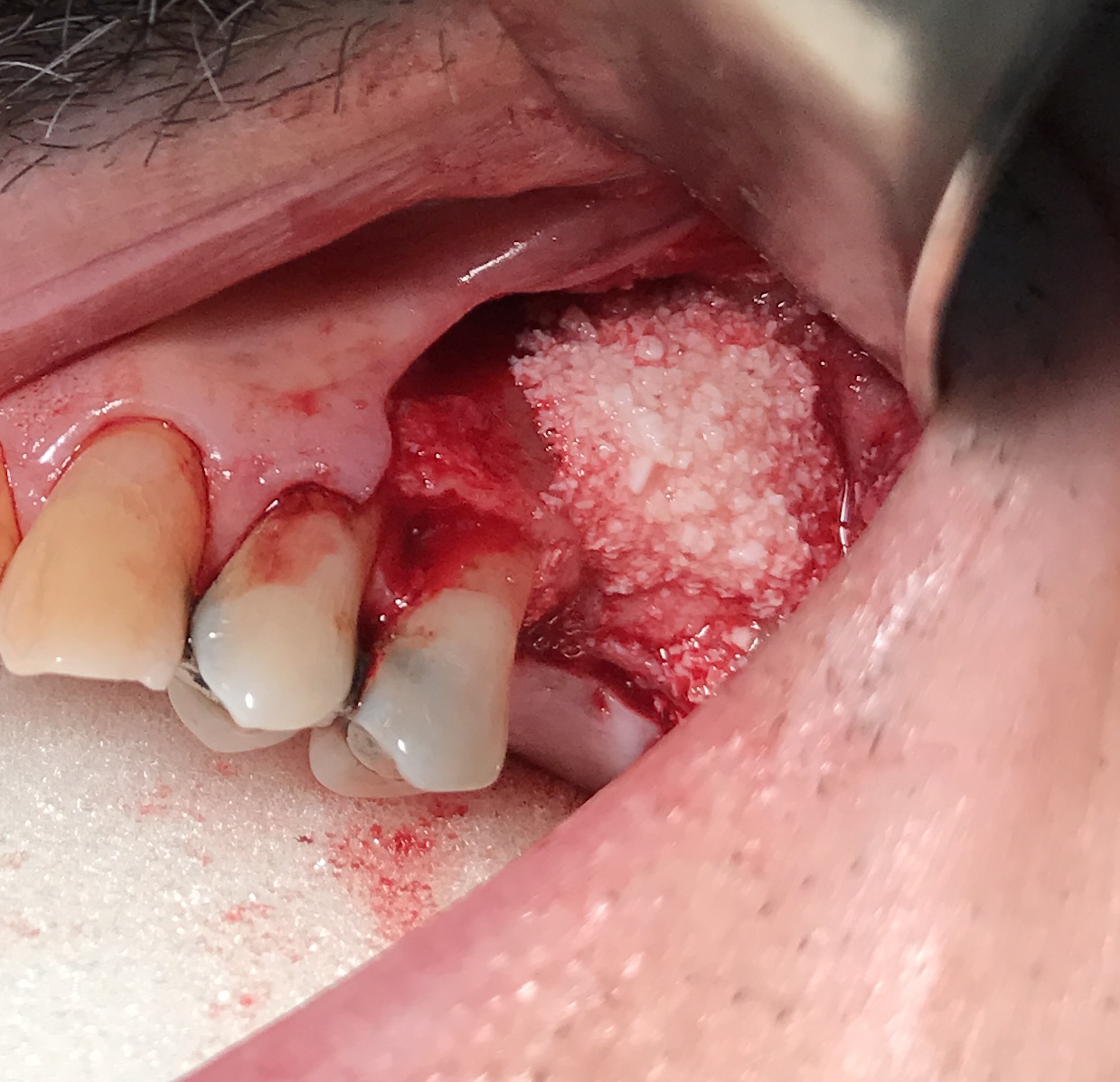
Bone Graft in place after Sinus membrane manipulation. Bone graft also used to increase the width of the bone.
PRF membranes covering the Bone graft.
Incision is closed. The Concave defect is now corrected by augmenting the defect laterally.
3D image shows newly augmented Sinus, and improved height of the bone in the area of maxillary left first molar.
by Abhishek Mogre DMD, Board Certified Oral and Maxillofacial Surgeon
"PRF" or Platelet rich fibrin is used in Oral Surgery for assisting in mucosal healing and faster angiogenesis and maturation of bone grafts. It is also used in third molar surgeries or "Wisdom Tooth" surgeries to reduce post surgical pain, and reduce the dependance on post surgical narcotic medications.
Following steps are involved in the preparation of PRF.
Collection of Venous "Autologous" (patient's own blood): In the picture below, Dr Abhishek Mogre is seen collecting patients own blood in a test tube (without any anticoagulant) using a 21g Vacutainer. The crucial step here is locate an appropriate peripheral vein, and the amount of blood required for the particular surgery has to be collected in less than 2 minutes. If the blood collection time increases, it will affect the quality of the PRF that is generated.
Transporting the collected blood and running the Centrifuge: As explained in the previous step, the tubes with collected blood have to placed in the centrifuge within 2 minutes of the start of the blood draw, and the centrifuge has to be run in a very specific setting. Dr Abhishek Mogre has chosen to use the original "Dr Choukran" concept and his centrifuge setting to generate the optimum quality of PRF product. It is very important to understand that the final product will vary if the centrifuge protocol is not followed. See the picture below of the centrifuge used by Dr Mogre in Shoreline Oral Facial Surgery & Dental Implants office.
Opening the caps of the test tubes and allow the PRF to self coagulate: Once the centrifuge has completed its appropriate cycle, remove the tubes, open the caps and let them stand in the test tube holder until the final product of PRF coagulates or "clots". It is important to understand that we are allowing "Physiologic" clotting process without any additives while preparing the PRF. The time for this step could vary between 5-15minutes.
Remove the 'PRF" from the test tube and place it in the PRF container for compression: The PRF is the middle layer in the test tube, and is gel like in consistency. It is carefully removed from the tube and placed in a PRF container and then compressed into flat membranes. During this process, the exudate rich in Fibronectin, Vitronectin, Stem cells and growth factors is collected in the bottom of the container. This exudate is used to hydrate any Allograft or "Cadaver Bone Graft" or Xenograft or "Bovine or Equine bone graft". See the pictures below, where Dr Mogre carefully removes the gel like PRF and then places in the container. The final product after compression looks like flat membranes.
Using the Final compressed PRF for surgical use: The final PRF membranes can now be used for variety of oral surgery procedures. Commonly they can be mixed with the bone graft materials, used in extraction sites/lower third molar or wisdom teeth sites etc. In the picture below Dr Mogre is mixing the PRF membrane with the bone graft material to improve the success of the grafted site.
by Dr Abhishek Mogre, Board Certified Oral & Maxillofacial Surgeon
Platelet Rich Fibrin commonly known as PRF is a second generation Platelet Growth Factor.
It is developed conveniently by using an appropriate Centrifuge to spin patients blood and divide it in 3 layers. The bottom layer after the spin, is composed primarily of Red Blood cells. The middle layer or PRF is composed of Platelets, Stem Cells and White Blood Cells held together in Fibrin network. The top layer is acellular plasma.
PRF is then separated and is prepared in variety of different ways as appropriate for the surgery.
PRF is different from the previous generation Platelet growth factors in the following ways:
1. It contains no additives such as Bovine Thrombin.
2. The PRF Gel which comprises of Fibrin, Platelets, White Blood Cells & Stem Cells is much more stable than all previous generation Platelet Growth Factors.
3. The Cytokines and Growth factors from PRF are released and available for 1-2 weeks compared to few hours or a day in previous generation Platelet Growth Factors.
4. The process of generating PRF is very simple, and requires a good peripheral IV access.