A Sinus Lift Surgery is a procedure performed in the upper jaw or "Maxilla" in the region directly adjacent to the Maxillary Sinus, commonly to augment/replace the missing bone required for Dental Implant placement.
After extraction of teeth close to the floor of the maxillary sinus, surgeon sometimes encounter lack of adequate bone to successfully place a Dental Implant to replace the missing teeth. This happens as a result of a combination of natural reduction in bone volume at the extraction site and an increase in the sinus volume.
In such cases, an Oral Surgeon performs a "Sinus Lift" surgery, to add bone in a space created by meticulously handling the sinus membrane.
This surgery can be performed under Local Anesthesia or under IV Sedation/General Anesthesia.
Bone graft requires 4-6 months of healing. Sometimes Dental Implants can be simultaneously placed during the sinus lift surgery, and your Oral Surgeon can discuss with you, about this option during the consultation.
Patients are placed on Antibiotics and Pain Medications after the surgery. Surgeons will provide specific post surgical instructions related to the sinuses. Pain and discomfort is usually well controlled and may last 48-72 hours. Some patients may experience increased sinus drainage in the immediate post surgical period.
The following pictures and video, will help in better understanding the surgical procedure. The surgery is performed by Dr Abhishek Mogre. The patient has reduced bone volume in upper left first molar, and was treated with Sinus Lift Surgery to replace the missing bone in preparation for a Dental Implant.
This 3D image shows lack of bone in the missing maxillary left first molar area and enlarged "Pneumatized" Maxillary sinus.
Bony defect in the Maxillary missing first molar area, seen as a concavity.
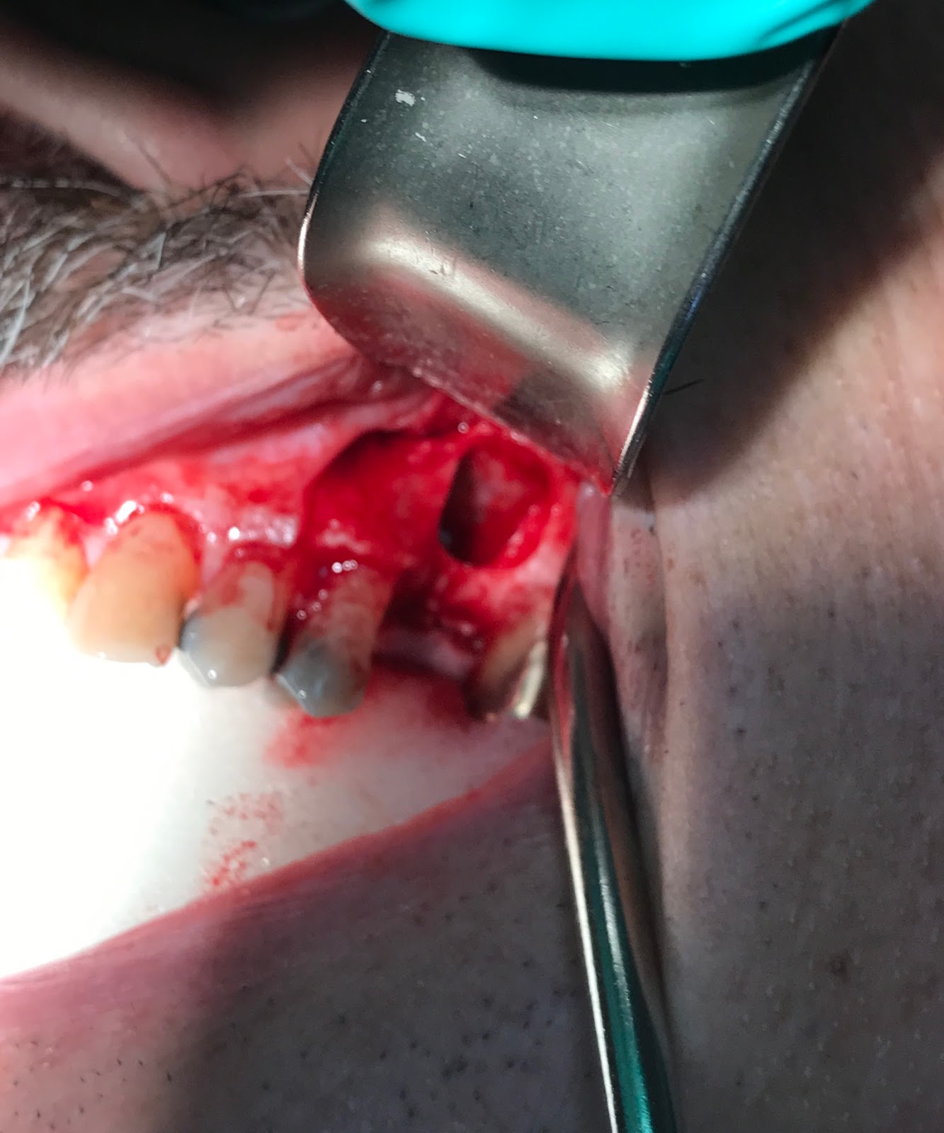
Surgical exposure of the defect
Access made to the Sinus Membrane with a window in the thin Bone in the area of the missing tooth. At this point, Sinus Membrane has been carefully manipulated.
The video shows movement of the manipulated sinus membrane during normal breathing.
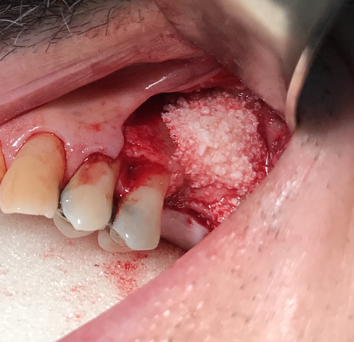
Bone Graft in place after Sinus membrane manipulation. Bone graft also used to increase the width of the bone.
PRF membranes covering the Bone graft.
Incision is closed. The Concave defect is now corrected by augmenting the defect laterally.
3D image shows newly augmented Sinus, and improved height of the bone in the area of maxillary left first molar.


Amazing piece of content, Thank you for sharing this blog....
ReplyDeleteOral Surgeon Pasadena, CA